Summary
A patient in her mid-50s complains of foot and leg pain. She’s post-menopausal with low bone density. A classic case of post-menopausal osteoporosis.
Not exactly.
And it won’t start to become clear until it gets to the point of her having repeated metatarsal bone fractures.
Let’s go back a little. It’s 2005. Our patient visits her family doctor complaining of pain in her legs and feet. But the discomfort she’s experiencing isn’t your typical aches and pains associated with aging.
“She develops a lot of [foot and leg] pain … So much so that she required pain management for this pain and her gate started to become affected,” shares Dr. Katherine Dahir, a professor of medicine at Vanderbilt University, who specializes in metabolic bone disease.
Her gait becomes wobbly and she’s experiencing an acceleration of degenerative changes in her spine. An osteoporosis screening reveals she has low bone density. She’s diagnosed with post-menopausal osteoporosis and is treated with bisphosphonates, the standard of care for patients with osteoporosis.
And this is when things get much more complicated.
Although all signs show an improvement in bone density, she begins to experience metatarsal bone fractures, which is highly unusual with osteoporosis. And not only does she have these unusual fractures, the fractures will not heal.
“And so that’s when you need to put your thinking cap on and try and figure out, why is this patient a treatment failure?” says Dr. Dahir.
To solve the case, the patient’s team studies her labs and finds a missing flag. “… It was called alkaline phosphatase, which is seen in a routine chemistry panel. Back at that time, it was only flagged if it was above the normal reference range because that usually indicates liver disease, but it wasn’t flagged if it was below the normal reference range because that was considered to be non-significant.”
But this finding would prove to be very significant. Combined with new research at the time, it helped identify a diagnosis for this patient — showing the importance of medical research that leads to more treatment options and more hope for patients.

Guest

Dr. Kathryn Dahir has been compensated by Alexion Pharmaceuticals, Inc. for her participation in partnership with The American Society for Bone and Mineral Research.
Dr. Kathryn Dahir is a Professor in the Department of Internal Medicine, Division of Endocrinology, Diabetes, and Metabolism at Vanderbilt University. She is the Director of Metabolic Bone Disease Program at VUMC and founding co-director of the Program for Metabolic Bone Disorders at Vanderbilt which focuses on pediatric to adult transitions of care for rare skeletal disease. Her effort is shared with clinical time devoted to care of patients with rare skeletal dysplasias including Hypophosphatasia, XLH, Osteogenesis Imperfecta, ENPP1 deficiency and Fibrous Dysplasia Ossificans Progressive as well as research program focusing on understanding the genetic basis of rare skeletal diseases. She is a member of multiple scientific advisory boards including the Magic Foundation, the International HPP registry, the OIF PCORI Scientific Advisory Board and the Soft Bones Scientific Board. Dr. Dahir has published more than numerous manuscripts in peer reviewed journals and authors the sclerosing bone chapter for Cecil’s Textbook of Medicine. She has won the highest patient experience award at Vanderbilt University Medical Center every year since 2017.
Transcript
DDx SEASON 6, EPISODE 4
Metatarsal Bone Fractures and a Rare Bone Disease Hiding in Plain Sight
RAJ: This season of DDx was produced in partnership with The American Society for Bone and Mineral Research, and sponsored by Alexion Pharmaceuticals, Inc.
Opening
RAJ: A woman in her mid-fifties complains of foot and leg pain.
She’s post-menopausal with low bone density.
Sounds like a bread and butter case of post-menopausal osteoporosis, right?
Not exactly.
Dr. Dahir: When I think about how best to describe this particular patient, really two phrases come to mind: one is hiding in plain sight, because this is a patient that honestly looked like any normal lady that you might see at the grocery store…Out of sight, out of mind, would be another good way to describe this case, because at that time, more than 15 years ago, her disease in adults was really not well described in the medical literature.
SHOW INTRO
RAJ: This is DDx, a podcast from Figure 1 about how doctors think.
I’m Dr. Raj Bhardwaj.
This season is all about rare bone diseases.
Today, a case from Dr. Katherine Dahir, a professor of medicine at Vanderbilt University, who specializes in metabolic bone disease.
Chapter 1: MISDIAGNOSIS
RAJ: It was 2005. Our patient saw her family doctor complaining of pain in her legs and feet.
But this wasn’t your typical aches and pains associated with aging.
Dr. Dahir: She develops a lot of pain. So foot pain and leg pain, and. So much so that she required pain management for this pain and her gate started to become affected.
RAJ: Up until now our patient had no mobility issues.
Dr. Dahir: But about the time in her mid fifties, she started to develop this wobbly gate and had a hard time getting from a chair to a standing position. And had acceleration of degenerative changes in her spine.
RAJ: A recent osteoporosis screening revealed she had low bone density.
Her family doctor diagnosed her with postmenopausal osteoporosis.
She’s treated with bisphosphonates, the standard of care when it comes to patients with osteoporosis.
And that is when things got much more mysterious.
Dr. Dahir: Even though all signs looked like she was improving in terms of her bone density, she started to break her feet.
RAJ: Patients with osteoporosis are prone to fragility fractures.
But multiple foot fractures… while on bisphosphonates… that’s unusual.
So her primary care provider referred her to Dr. Dahir.
Dr. Dahir: She was in her mid fifties and came to me for evaluation of osteoporosis. And within that first year, she developed multiple fractures of the metatarsal bones in both the right and the left feet and in that first year had five fractures. And so not only fractures that came on with no trauma, but they wouldn’t heal. And so that was something that was different and once she got that first fracture, the frequency of her fractures really escalated fairly quickly.
RAJ: So let’s review: our patient had leg and foot pain. Low bone density. Her mobility was affected, her gait was wobbly. And this was followed by many fractures that refused to heal.
Dr. Dahir: But the symptoms of the fractures and the pain and mobility were never put together potentially being part of the same disease.
RAJ: When she was treated with bisphosphonates, her bone density appeared to improve, but her fractures became worse. Another significant clue.
Dr. Dahir: So she is the lady that we think we have on appropriate therapy for osteoporosis, but rather than preventing fractures, her fractures seem to accelerate. And so that’s when you need to put your thinking cap on and try and figure out… Why is this patient a treatment failure?
Chapter 2: DIAGNOSIS
RAJ: In her clinic Dr. Dahir reviewed the patient’s labs.
Dr. Dahir: One of the things that you do in your detective work is you look at the biochemistry or you look at the labs that were checked and historically going back. All of her labs were normal.
RAJ: But one result gave her pause.
Dr. Dahir: She had one specific lab that wasn’t flagged, it was called alkaline phosphatase, which is seen in a routine chemistry panel. Back at that time, it was only flagged if it was above the normal reference range because that usually indicates liver disease, but it wasn’t flagged if it was below the normal reference range because that was considered to be non-significant.
RAJ: But in this case it turned out to be very significant.
Dr. Dahir: Going back through and trying to figure out why is this person fracturing, even though she’s compliant, her bone density looks better, her labs look better, and the only thing to go on at that point was this low alkaline phosphatase. What was in the medical literature at the time is that a low alkaline phosphatase was associated with an ultra rare autosomal recessive disease called hypophosphatasia.
RAJ: Hypophosphatasia is a rare genetic disease that prevents the mineralization of bone. Bones become soft and fracture easily. It can be mild, moderate or deadly and occur at any age. Its symptoms are wide-ranging. Someone with hypophosphatasia might have short bowed limbs, knock knees, unexpected tooth loss or other bone deformities.
But our patient just didn’t look like someone with this condition.
Dr. Dahir: So what was really interesting to think about, wow, this lady has fractures and she has a low alcohol and phosphatase, but she doesn’t look like she has a severe skeletal dysplasia. She doesn’t look like these babies that are described in the medical literature.
RAJ: There was only one way to know for sure.
Dr. Dahir: And at that time, since we’re talking about more than a decade ago, it was really hard to get genetic testing because it wasn’t routinely covered by insurance. And I sent genetic testing thinking, could this lady potentially have the same gene as some of these babies that have the autosomal recessive form in the literature? And back then when you sent genetic testing, you had to send it and forget about it for a while because it would take a long time before it would come back.
RAJ: While waiting for the results of the genetic testing, Dr. Dahir continued her evaluation.
She needed to know whether this patient suffered from osteomalacia, a decreased mineralization of bone, and a key symptom of Hypophosphatasia.
So she requested a bone biopsy to find out.
Dr. Dahir: This is how it works when a patient gets a bone biopsy that you send them to the orthopedic surgeon and they go to the operating room and they go in with a little needle that’s called a trocar, and they take a little piece of bone from the anterior iliac spine. So that’s the part of the pelvis and they’re able to take this piece of bone and then it gets analyzed and you can do metabolic studies and see how much of that bone is mineralized versus unmineralized.
RAJ: The next day, Dr. Dahir got an alarming phone call from her patient.
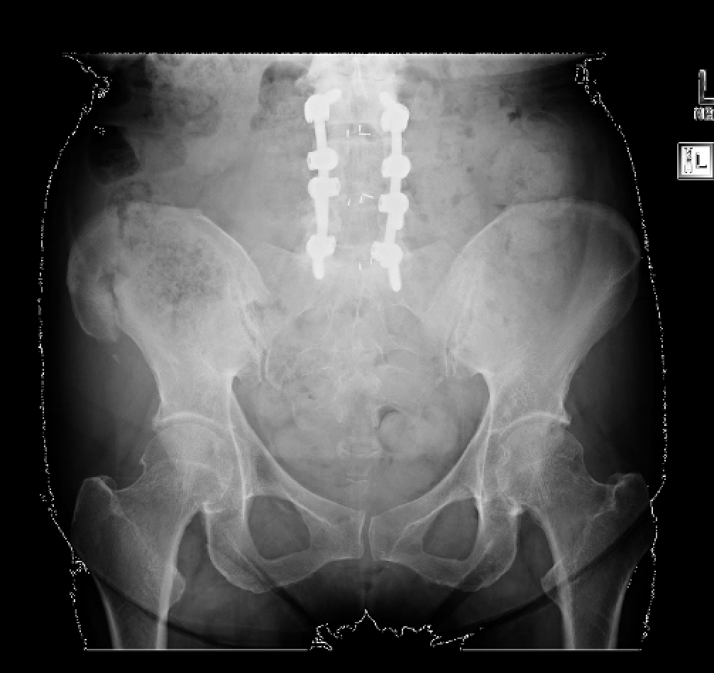
Dr. Dahir: She calls me and she says, “I’m having so much pain, I can’t walk.” So I sent her to the emergency room for pelvis films and I couldn’t even believe it. Her entire sacrum had fractured just due to this tiny needle getting put, in the operating room under anesthesia with the best standard of care, a thoughtful orthopedic surgeon doing this, her x-ray of her pelvis with that little trauma with the needle. The radiologist actually called me and said, This lady looks like she was in a motor vehicle accident. Her sacrum had fractured completely through. And that was really scary for me to see for this patient, her bone was so fragile that a tiny bit of trauma caused that pelvis to just break like that.
RAJ: If her fractured sacrum wasn’t enough evidence… the biopsy confirmed that her bones were dangerously fragile.
Dr. Dahir: And as you would expect when we got her bone biopsy results back there was very little mineralization. So the bone of that biopsy site was just this unmineralized connective tissue or collagen. And to put that into context, you know what part of our body is just collagen and it’s not mineralized, it’s like the tip of your nose. So that doesn’t feel very strong. Think about if you were walking around on that, and that was the quality of her bones?
RAJ: But what about the timing of her fractures? Was it significant that they increased after our patient was put on bisphosphonates?
As it turned out, it absolutely was.
Dr. Dahir: She was on a medicine called a bisphosphonate for osteoporosis that can actually make this situation worse because it’s what’s called APY phosphate analog. It can unmask symptoms of hypo phosphate.
RAJ: A few weeks later, the genetic test results were in.
Dr. Dahir: She had not two affected genes associated with hypo phos. She had one, so she looked to be what at the time we would have thought of a carrier of hypo phosphate. But she really manifested the disease, but not so much as a baby, but as an adult.
RAJ: At that time in 2012, The New England Journal of Medicine had published a study about an enzyme replacement therapy which was a breakthrough treatment for infants with Hypophosphatasia.
But the study also highlighted something significant about our patient.
Dr. Dahir: Looking at her particular gene, it was one of the genes described in that New England Journal and medicine paper in tandem with a separate affected gene. And so my eureka moment was wow this disease can present in a milder form later in life with just one gene, so it can be transmitted both autosomal recessive as well as autosomal dominance. And once we had that information, we were able to get her on the right path for treatments.
Chapter 3: LESSONS
At the time, our patient’s case was fairly unique. But today much more is understood about hypophosphatasia in adults.
Dr. Dahir: The good news is that there’s so much more in the medical literature describing milder forms that hopefully it’s not such a scary diagnosis for patients to read about when they’re doing their own research. And we do have treatment options that are available now. Plus, there are newer treatment options in the clinical trials pipeline as well so more hope for patients.
SHOW CLOSING
RAJ: Thanks to Dr. Dahir for speaking with us.
This is DDx, a podcast by Figure 1.
Figure 1 is an app that lets doctors share clinical images and knowledge about difficult to diagnose cases.
I’m Dr. Raj Bhardwaj, host and story editor of DDx.
You can follow me on Twitter at Raj BhardwajMD.
Head over to figure one dot com slash ddx, where you can find full show notes, speaker bios and photos – including the x-ray of our patient’s fractured sacrum.
This episode of DDx was produced in partnership with The American Society for Bone and Mineral Research and sponsored by Alexion Pharmaceuticals, Inc.
For more information on hypophosphatasia please visit Alexion.com
Thanks for listening.


