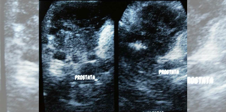
A 61-year-old presents with elevated PSA levels during routine screening. Digital rectal examination reveals palpable abnormalities in the prostate gland. Further investigation with MRI indicates the presence of a suspicious tumor within the prostate gland. Given these findings, a biopsy is performed to confirm the diagnosis.
Clinical Findings
The biopsy results confirm the presence of prostate cancer. Histological examination reveals characteristic features typical of prostate adenocarcinoma. The histopathological findings include a Gleason score of 7 (3+4), suggesting intermediate aggressiveness grade cancer. Overall, imaging and histological findings did not provide evidence of cancer cells spreading outside the prostate gland.
Treatment
Since the patient is of relatively young age and evidence supports localized prostate cancer, the patient undergoes a radical prostatectomy procedure utilizing robotic-assisted technology. This approach is chosen to minimize the risk of side effects and preserve nerve function, in order to minimize the risk of urinary continence and sexual dysfunction post-surgery.
Postoperative care includes continuous monitoring of PSA levels to track any potential recurrence of cancer. Additionally, the patient undergoes physiotherapy to mitigate the risk of urinary incontinence commonly associated with prostatectomy procedures.
Ongoing Management
Regular follow-up appointments are scheduled to assess the patient’s recovery, check PSA levels, and review any potential complications that might arise.
References
Seo, Hyun-Ju, et al. “Comparison of robot-assisted radical prostatectomy and open radical prostatectomy outcomes: a systematic review and meta-analysis.” Yonsei medical journal 57.5 (2016): 1165.
Figure 1 – The future of medical education [Internet]. [cited 2024 Sept 4]. Comprehensive Approach to Prevention and Management of Prostate Health. Available from: https://app.figure1.com/cme/9c5587f2-9976-40be-8eac-c338d8df291e
Figure 1 Clinical Case: https://app.figure1.com/cases/306cbc1f-b1af-434c-acad-8f4047de9e50
Published September 4, 2024
Want more clinical cases?
Join Figure 1 for free and start securely collaborating with other verified healthcare professionals on more than 100,000 real-world medical cases just like this one.