Healthcare professionals regularly share new and fascinating cases to Figure 1. Here are five of the most interesting medical cases of Crohn disease and colitis causing buzz in the community.
#5.
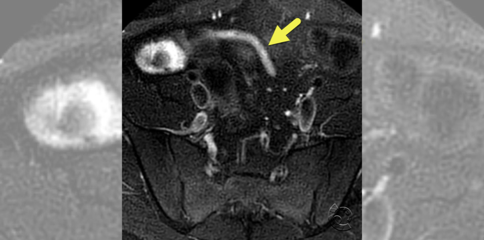
This MRI image shows mild inflammation of the appendix (arrow) in a patient with Crohn disease. According to the Figure 1 member who shared this case, “The appendix can be inflamed in patients with Crohn disease. Isolated Crohn appendicitis is rare, occurring in less than 1% of patients with Crohn disease. Patient is often present with signs of acute appendicitis.”
#4.
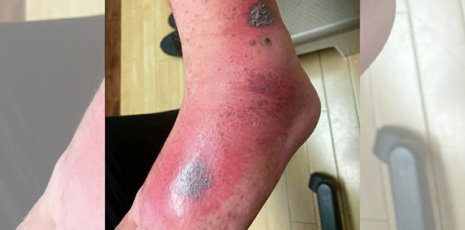
An infectious disease physician shared the following details of the fourth most interesting medical case of Crohn disease and colitis: “Subacute progressive lesions … involving bilateral lower extremities (only shins and knees). The initial onset of multiple tender ulcerative lesions (1-2cm) began a few weeks prior while hiking in Germany, Denmark and Norway. He was careful about checking for ticks, no contact with animals (except once petting a stray cat), and stayed in Airbnbs, used jacuzzi once. Never had fever or other associated symptoms, basic labs were normal, and he did not respond to amoxicillin, clindamycin, steroids while in Germany (presumed erythema nodosum). The dark lesions (at various stages of progression) expanded and became more ulcerative, erythematous and painful until he presented for admission back in the US. PMHx: managed for #Crohns with 6MP and #adalimumab w/o a recent flare for ~13 years.”
The patient was then admitted “for IV treatment of underlying cellulitis with vancomycin and the surrounding pain/erythema improved (photo #4). CRP: 10. ESR: 24. Fungitel neg, TB Quantiferon neg, HIV neg, HBV neg, histo Ag neg, anti-adalimumab Ab neg. A punch biopsy was sent from right ankle for fresh tissue culture, which had no growth, no AFB; and separate sample for path, which revealed …”
#3.
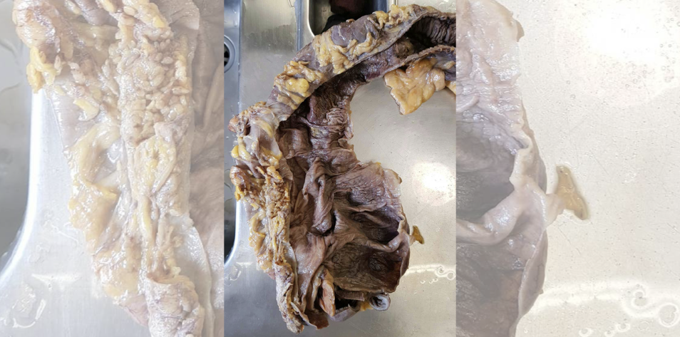
This image shows a “whole colon removed for pseudomembranous colitis in a patient with … early carcinoma of the rectum,” according to the pathology resident who shared the case with the community.
A registered nurse asked about why the colon appeared to be gray and a technologist shared the following: “It normally would be [pink in color] but after resection has taken place the specimen has no blood supply and is also preserved usually in formaldehyde” which causes the gray coloring.
See all the medical case images
#2.
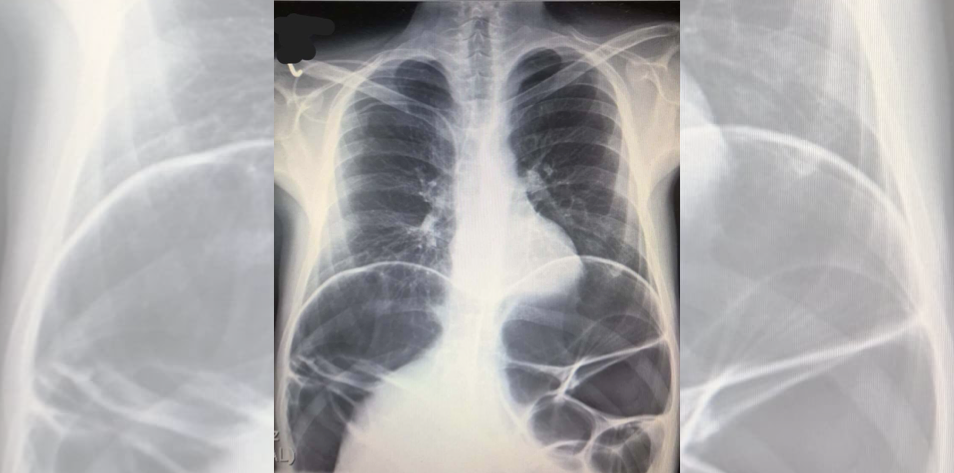
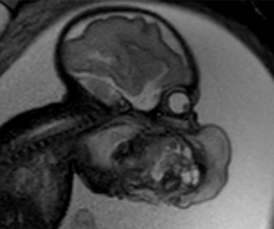
This image shows a megacolon in a patient with Crohn disease. The gastroenterologist who shared the case noted that the patient originally came to the emergency department after stopping azathioprine and a steroid medication that caused abdominal issues. The patient required surgical intervention, which is when the “fistula to the urine bladder was found.”
According to the gastroenterologist, “There were two obstructions, one between the rectum and the sigmoideum, and one in the terminal ileum. The obstructions and the rectovesical fistula were removed, now he has an [ileostomy] for a few weeks, then he will be reoperated. It’s strange, but his urine tests were all negative.”
#1.

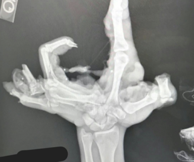
In this most interesting medical case of Crohn disease and colitis, a patient with diabetes presented to the emergency department for an infection. During physical assessment, the leg wounds, which were not part of the original complaint, were discovered.
According to the registered nurse who shared the case, “He stated [the wounds] had been there for years and nothing makes them better — had never tried any formal medical treatment. Ulcers were so deep he had arterial erosion and bleeding twice, once requiring cautery. After a couple days and a wound consult, the patient shares that he once was told he had ulcerative colitis.”
Published July 15, 2024
Join the Conversation
Sign up for Figure 1 and be part of a global community of healthcare professionals gaining medical knowledge, securely sharing real patient cases, and improving outcomes.