Healthcare professionals regularly share new and fascinating cases to Figure 1. Here are five of the most interesting medical cases of teratomas causing buzz in the community.
#5.
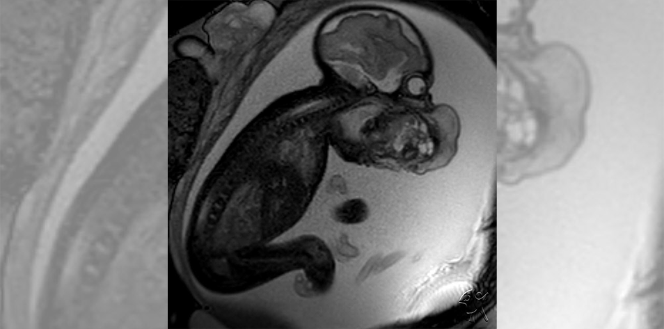
In this fifth most interesting medical case of teratomas, a fetal MRI shows “an epignathus, or a teratoma arising from the oropharynx. Because of the location of the tumor, the fetus has difficulty swallowing leading to polyhydramnios. The tumor is identified on prenatal imaging as a mass extending out of the mouth. The fetus has a poor prognosis due to airway obstruction. Depending on the location, size of the tumor, and associated anomalies, the fetus can be delivered via an ex utero intrapartum treatment (EXIT) to tracheostomy procedure.”
The procedure is described as a partial delivery, where, while still attached to the placenta via the umbilical cord, a surgical team “attempts to secure an airway”.
Commenters on the case noted the difficulty of the procedure with one pulmonologist sharing, “This is a very delicate case, EXIT being a possible option is yet associated with so many complications. Bleeding is a very much anticipated one, for tracheostomy to be done, the mass is even almost occupying the whole of the neck, considering neovascularization of the mass. Edema is another, etc.”
#4.
This medical case details an 18-year-old who has never been pregnant reporting hypogastric and abdominal pain. The OBGYN who shared the case included these details: “Her condition started a month prior to consult. Her menstrual period was normal, regular. Transrectal ultrasound revealed that the right ovary was converted into a multiloculated cystic structure measuring 27x22x14 cm with papillary projections and minimal color flow studies. She was requested for tumor markers and further work up however patient complained of persistent pain with pain scale of 8/10.”
While in surgery, a ovarian cyst was discovered with “one full twist on its pedicle.” Histopathy revealed the final diagnosis.
#3.
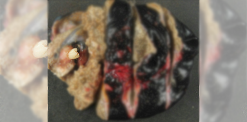
This third most interesting medical case of teratomas shows a mature cystic teratoma from a patient with “bilateral ovarian teratomas,” according to the pathology resident who shared the case. The resident noted, “The diameter was approximately 3-4 inches and the outside is actually inked black, so it didn’t originally look like that.”
Regarding the tooth that had formed, the resident gave this explanation: “Mature cystic teratomas are benign tumors (no metastatic potential) derived from pluripotent cells (in this case germ cells of the ovary, but can be from embryonal cells occurring anywhere in the midline of the body such as the neck or coccyx). B/c the cells from which they are derived are pluripotent, they may form a teratoma from one or more of the three developmental germ layers. The three primary germ layers are ectoderm, mesoderm, and endoderm. The tooth is formed from the ectoderm, while some other components of the teratoma may form from mesoderm (cartilage, muscle) and/or endoderm (thyroid, GI tract).”
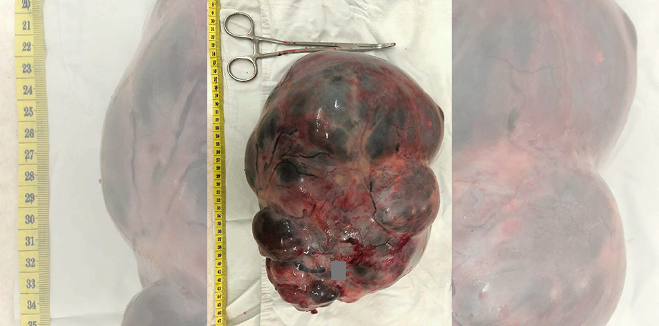
#2.
In this case we see the cross section of a teratoma that formed in a 35-year-old patient near the retroperitoneal, almost near the spleen. Commenters were fascinated by the case image, commenting, “This image made my night! I always wanna see whats inside!” and “Impressive!”.
#1.
In the most interesting case of a teratoma, this teratoma was discovered in a 17-year-old who, according to the registered nurse who shared the case, “was having LLQ pain, but the surgeon had no idea, until he got in, that there was a #Teratoma and to what extent. This one had a LOT of hair and some teeth.”
Published September 16, 2024
Join the Conversation
Sign up for Figure 1 and be part of a global community of healthcare professionals gaining medical knowledge, securely sharing real patient cases, and improving outcomes.